Use of laboratory animals to identify carcinogenic potential of chemicals, mixtures, and other agents has a modern history of greater than 40 years from which much useful scientific and public health information can be derived. While laboratory animals differ from humans in some respects that may affect responses to hazardous exposures, use of such models is based on experimental evidence indicating that there are more genetic, genomic, physiological, biochemical, and metabolic similarities than differences among mammalian species. Issues of concordance of responses between rodent species and between rodents and humans as well as repeatability and site-specificity are important considerations in evaluating laboratory animal carcinogenicity results. Variables in experimental design such as animal strain, diet, route of exposure, and study duration as well as single-site versus multisite carcinogenic responses all influence interpretation and intelligent use of study data. Similarities and differences in site-specific laboratory animal and corresponding human cancers should also be considered in study evaluation. Recent attempts to explore genetically engineered mice and to humanize the mouse for more relevant identification of carcinogen hazard identification have yielded mixed results. In the end we are confronted by the realization that virtually all animal cancer models are useful but imperfect surrogates for humans. Assuming the percentage of chemicals currently in commerce that are estimated to be potent animal or human carcinogens is quite low, the task of identifying agents with significant carcinogenic potential is daunting and important. The biological conundrum of scientific debate regarding the relevance of carcinogenicity studies in laboratory animals is likely to continue. Nonetheless public health considerations must take precedence when deciding human safety issues.
Keywords: Cancer bioassays; carcinogenesis; species differences; comparative pathology; hepatocarcinogenesis; genetically engineered mice; cancer prediction
INTRODUCTION
Historically, laboratory animal studies, especially studies in mice and rats, have made highly tangible contributions to physiology, biochemistry, pharmacology, medicine, toxicology, and cancer biology. Use of rodent models is based on the premise and supporting experimental evidence that rodents have sufficient physiological, biochemical, metabolic, and genetic/genomic similarities to humans to warrant their use as surrogates, as well as on the basis of their relative availability, low cost, and short life span. These studies have been and continue to be fundamental for the discovery of new disease treatment regimens and beneficial chemicals and to help determine the efficacy of new pharmaceuticals and anticancer therapies. Preclinical studies in laboratory animals typically precede human clinical trials and population studies for new drugs and therapeutics. Laboratory animal studies contribute to our understanding of mechanisms, modulators, and pathogenesis of disease, study of specific diseases, discovery of the underpinnings of cancer, and evaluation of intervention strategies. Similarly, and most importantly, they allow us to identify potential public health hazards.
Use of rodent models to identify carcinogenic potential of chemicals and other agents has a long and eventful history. The chemical carcinogenesis revolution and testing age began when Yamagiwa and Ichikawa in 1918 showed that coal tar experimentally applied to rabbit ears caused skin carcinomas (Yamagiwa and Ichikawa, 1918). Since then and especially over the past 4–5 decades, we have gained considerable insight into the strengths and weaknesses of toxicity and carcinogenicity studies in laboratory rats and mice. The 2 largest, longest existing, and most well established bioassay programs in the world are the Ramazzini Foundation (RF) and the National Toxicology Program (NTP) (Huff, 2002; Soffritti et al., 2002). Together more than 700 chemicals or agents have been tested for carcinogenic activity by these two programs: roughly 200 by RF and 500 by the NTP.
Over the years, and especially in the last decade, there has been considerable debate regarding the value of rats and more particularly mice in testing agents for carcinogenic potential (Schach von Wittenau and Estes, 1983; Monro, 1993; Huff, 1994; 1999; Johnson, 2003; Wagner, 2003). What has emerged from this debate is a better appreciation that the interpretation and use of results from rodent carcinogenicity studies should take into account possible modulators of the observed responses and awareness of the experimental exposure conditions in some rodent studies that might not be anticipated to occur exactly the same in humans (Monro, 1993; Cohen, 1995; Abdo and Kari, 1996). Thus, extrapolations from a single rodent study to humans may be subject to potential errors (Purchase, 1980).
In the past, criticism of the use of rodent bioassays for identification of carcinogenic potential has focused on concerns about positive findings in rodents considered not to be relevant to humans. What seems to have been overlooked is that no one ever said that rodent carcinogenicity bioassays were perfect surrogates for humans. Nonetheless, we do know that for now and until something better, more rapid, less expensive, and more accurate and predictable is found, long-term chemical carcinogenesis bioassays remain the best and most globally accepted means we have for identifying potential human carcinogens (Tomatis et al., 1997; 2001).
Value of Animal Experiments
No reasonable scientist would claim that rodents are perfect surrogates for humans. In fact, rats are not perfect surrogates for mice, and vice versa, but for carcinogenicity the correlation is beyond 74% (Haseman and Huff, 1987). This is precisely the reason that bioassays include both rats and mice—because their sensitivities for different chemicals or different classes of chemicals may be complementary. Carcinogenic potential, not absolute identification of human carcinogens, is being measured. Although we know that all known human carcinogens are also carcinogenic to rodents, it is noteworthy that nearly one-third of these were first identified in animals and only subsequently in humans (Tomatis, 1979; Huff, 1993b).
Outcomes of any study are influenced by multiple factors (e.g., experimental design, exposure levels, species, strain, sex, route of exposure, duration of exposure, metabolism, diet, and pathology) (Haseman, 1983; Schut et al., 1983; Gregory, 1988; Wolff et al., 1991; Griesemer and Eustis, 1994; Festing, 1995; Ginsberg et al., 1996; Dass et al., 1998; Keenan et al., 1999; Lovell et al., 1999). Although not perfect, there is enough concordance between human and rodent carcinogens, in repeatability of bioassay results, and in site-specificity to warrant continued use of existing hazard identification testing approaches until such time as we develop a more suitable means of identifying agents with human carcinogenic potential. At present, the rodent cancer bioassay is the best tool available, provided we are mindful to effectively deal with and interpret false-positives, false-negatives, metabolic differences between species, and species-specific responses that will likely occur.
The advent of genetically engineered mice and sophisticated gene-transfer technologies, including the ability to turn on and off specific genes, promises to permit the development of models with multiple genetic alterations to more closely mimic the multiple complex aspects of carcinogenesis (Marx, 2003). Hopefully, biomarkers will be found that will identify cancer risk early enough for intervention strategies to be effective. Progress in exploring the utility of genetically modified mice in cancer hazard identification is underway (ILSI/HESI, 2001; Pritchard et al., 2003) and development of genetically engineered models with specific alterations believed to be important in human cancer is being funded by National Institutes of Health consortia (http://emice.nci.nih.gov/); (http://www.niehs.nih.gov/cmgcc/home.htm).
Species Differences and Similarities in Carcinogenesis
Although differences may exist in the morphologic features, pathogenesis, and molecular and cellular processes of carcinogenesis between species, considerable similarities are found to warrant utilization of animal models to understand certain aspects of the process and to identify agents with carcinogenic potential. It is a given that we do not have a complete understanding of etiology, pathogenesis, and natural history of neoplasia. With respect to chemically induced cancer development, the heterogeneity of target tissue response within a given species is as great as the heterogeneity between species or sexes.
Despite these differences between species, there are important similarities favoring continued use of animal models. “Experimental evidence . . indicates. . that there are more physiologic, biochemical, and metabolic similarities between laboratory animals and humans than there are differences. These similarities increase the probability that results observed in a laboratory setting will predict similar results for humans. Clearly the accumulated experience in the field of carcinogenesis supports this concept.” (Rall et al., 1987)
Because rodents appear to be more susceptible to development of cancer in a wider variety of tissues (Grisham, 1996), in contrast to humans, doesn’t make them less valuable as research and testing tools. There is credence, however, to the notion that if humans underwent as complete pathology/histopathology as do rodents, the cancer gap would be substantially reduced or even eliminated. In fact, their utility as experimental models and test species to identify carcinogenic potential is favorably enhanced by the increased numbers of tissues/organs evaluated histopathologically. Differential susceptibility among rodent strains to chemical carcinogenesis may be related to a variety of factors such as gene imprinting and the time required for development of cancer (Haseman et al., 2001). Researchers and public health investigators can capitalize on this differential susceptibility in their respective studies.
In contrast to observations in humans, rodents utilized in cancer bioassays frequently develop multiple neoplasms at different tissue sites. In almost all cases in which human carcinogens have been tested in animals, there is at least one or more common target sites for carcinogenesis (Huff, 1994; Tomatis et al., 1989; Wilbourn et al., 1986). The exact reasons for this are unknown but relative genomic instability, differences in genomic imprinting, cancer susceptibility loci, and the possibility that higher dose levels used in rodent cancer bioassays may impact the carcinogenic response are possible explanations. Of course many of these higher exposure levels are similar to occupational exposures (e.g., benzene, butadiene, methylene chloride, tetranitromethane), especially those in the past, and to contemporary cancer chemotherapeutic exposures (Freireich et al., 1966).
Rodents also sometimes develop neoplasia at tissue sites that are unique, i.e., there is not an exact morphologic counterpart in humans. Examples include Zymbal’s glands, forestomach, Harderian gland, and preputial gland (Huff, 1992). Although neoplasms in these rodent-unique tissues could not occur in humans, this does not make them irrelevant. For example, the first identified cancer in rodents for the leukemogen benzene was the Zymbal gland (Maltoni and Scarnato, 1979). Furthermore, since neoplasia at these specific sites does not occur universally in rodent cancer bioassays, when present they certainly represent a carcinogenic response and must not be dismissed as irrelevant. The tissue components in Zymbal’s gland, forestomach, Harderian gland, and preputial gland correspond to similar cells at other sites in humans. Cellular and molecular changes leading to cancer at these sites are expected to be similar to cellular and molecular changes seen in human cancers. Mechanistically it matters little what the eventual cancer target site may be; the important observation is whether a chemical does or does not cause cancer.
Comparative Anatomy, Histology, and Neoplastic Pathology of Humans and Rodents
Comparative Anatomy: Despite the obvious differences in scale, the internal murine anatomy is remarkably similar to that of humans. In fact, rodents possess all of the major organs possessed by humans in the cardiovascular, respiratory, nervous, digestive, endocrine, and genitourinary systems. The gross anatomy of the individual organs of these systems is also basically similar to that of humans. There are minor differences from humans in the structural organization of some rodent organs, such as the number of right and left lung lobes, the bicornuate uterus, and the division of the prostate into separate lobes. And there are differences in the location and number of some rodent organs, such as the contiguous alignment of the three major salivary glands and the presence of several pairs of mammary glands (five in mice, six in rats) extending from the ear to the base of the tail, but the basic anatomy of these glands is similar to that of humans.
In addition to possessing all of the major organs of humans, rodents have three glands without an obvious human counterpart—the intraorbital Harderian gland, the male preputial gland, and the female clitoral gland. Rodents also have an auditory sebaceous gland (Zymbal’s gland) in the external ear; however, this gland is not entirely unique since the skin of the external auditory meatus of humans contains hairs with sebaceous glands that bear a morphologic resemblance to the Zymbal’s gland. Finally, there is the rodent forestomach with a mucosal lining similar to that of the human esophagus but occupying the first portion of the gastric pouch and lacking in humans. Since these additional glands and forestomach of rodents have no direct human analogue, it might be argued that tumors arising in these organs as the result of chemical induction are inconsequential with regard to human risk. On the other hand, these structures could be regarded as bonus organs, which serve as additional bellwethers of a chemical’s carcinogenic potential whenever tumors also develop in one or more vital rodent organs with comparable human analogues.
Comparative Histology: As might be expected from the anatomical similarities, the histologic features of most rodent organs are roughly similar to those of humans. Nonetheless, minor differences may be noted in the histology of many organs, such as the relatively large adrenal medulla of rodents in comparison to the thickness of the adrenal cortex, the striking prominence of the Clara cells in murine bronchioles, the cuboidal parietal epithelium in the Bowman’s capsule of male mice, and the relatively greater fat/glandular tissue ratio in the mammary glands of rodents. Furthermore, two fundamental differences that seem to apply to most rodent organs and tissues are the less abundant interstitial connective tissue in comparison to human tissues, and the less abundant smooth muscle in a variety of tissues such as the gastrointestinal tract, the uterus, and the prostate. The muscular wall of the intestinal tract is particularly thin in rodents in comparison to the thickness of the intestinal mucosa, and the prostate gland of rodents contains very little smooth muscle between the glandular elements in contrast to the human prostate gland in which smooth muscle comprises 30% of the total mass. The impact of these differences in mesenchymal elements is relatively minor in regard to comparative neoplasia since malignant neoplasms arise in the connective tissues and smooth muscle of rodents and humans relatively infrequently. A minor consequence, from the pathologist’s perspective, is that the presence or absence of tumor invasion may be more difficult to evaluate in rodents because there is so little surrounding or supporting mesenchymal tissue.
Comparative Neoplastic Pathology: In view of the similar anatomy and histology, one might expect the incidence of neoplasia, as well as the prevalence of histopathologic types, to be similar as well. If the natural incidence or specific histopathology differs significantly, one might infer that the rodent may not be a suitable surrogate model for humans. To reach this conclusion, one would have to believe that neoplasia is solely dependent upon the genetics of an organism, and that the environment plays no role. If this were so, then the incidence of human neoplasia might be only one-tenth of the actual current incidence, and the impact of neoplasia in humans would be significantly reduced. Of course, we now know that environmental factors play a major role in many types of human cancer, and perhaps none of these factors is more important than that of cigarette smoking. In fact, it has been estimated that 30% of all cancer deaths are attributable to cigarette smoking.
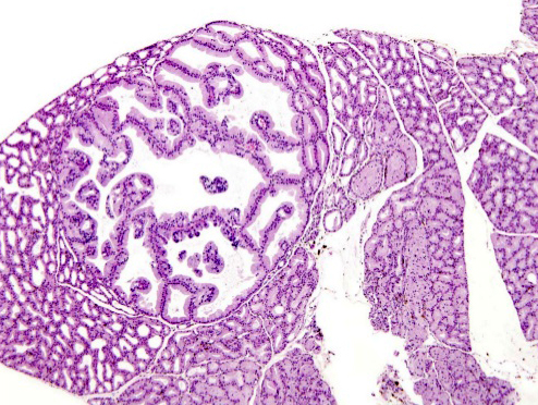
The point is that rodents are not exposed to the same environmental carcinogens as humans, unless they are artificially exposed in the laboratory, and therefore they would not be expected to have the same incidence of neoplasia or the same neoplastic histopathology in every organ. For instance, in rodents, tumors arise relatively infrequently in the pancreas and most commonly involve the acinar elements, resulting in acinar cell adenomas and carcinomas (Eustis and Boorman, 1990). On the other hand, pancreatic cancer in humans is the fifth most common cause of cancer related death, and 90% of the time the cancer involves the ductal elements, while neoplastic disease of the acinar elements is rare (Scarpelli, 1988). Does this mean that the rodent is a poor model? In fact, these species differences in incidence and histopathology may be explainable in part by environmental exposures in humans since cigarette smoking is the most firmly established risk factor in pancreatic cancer, and the chronic pancreatitis resulting from the abuse of ethyl alcohol is a risk factor as well.
To further examine the role of environmental factors in explaining neoplastic differences between species, it is instructive to compare the major morphologic categories of pulmonary neoplasia, since tumors of the lung are common in both rodents and humans. In rodents the predominant form of neoplastic lung disease is of the alveolar/bronchiolar type. This is true of both mice and rats, and it is also characteristic of both spontaneous neoplasia and chemically induced increases in neoplasia, although squamous carcinomas can be induced with certain chemical carcinogens. In humans, on the other hand, the predominant forms of pulmonary cancer are adenocarcinoma, squamous carcinoma, and undifferentiated small and large cell carcinomas, with bronchioloalveolar carcinoma representing only a minor percentage of the total, ranging from 1 to 9% in various series.
To some extent, the differences in incidence of bronchioloalveolar carcinoma between the species may be explained by the manner in which the tumor is defined. That is, in humans there is a very restrictive definition that limits the use of the term to those tumors with a lepidic growth pattern and absence of any invasion of stroma, vessels, or pleura. Thus, many of the carcinomas of the lung in rodents would probably be classified as papillary adenocarcinomas in humans because of the presence of invasion. A more accurate comparison, then, might be to combine all of the adenocarcinomas and bronchioloalveolar carcinomas in humans, and the resulting difference in incidence from that of rodents would be significantly reduced.
There is a more important factor than the definition of morphologic types, however. And that factor, of course, is cigarette smoking, which is the cause of the vast majority of lung cancer in humans. The increased risk in smokers is seen for all types of lung carcinoma, but the percentage of smokers is particularly impressive for the major subtypes of carcinoma: small cell carcinoma (98.9%), squamous cell carcinoma (98.0%), large cell carcinoma (93.3%), and adenocarcinoma (81.6%) (AFIP, 1995). Although smoking is also a risk factor for bronchioloalveolar carcinoma, the percentage of smokers among those having this disease is the lowest of all major types (70.6%). If we now consider the morphologic subtypes that might remain if smoking could be eliminated as a factor, the data would suggest that the great majority of squamous cell carcinomas, small and large cell carcinomas, and 80% of the adenocarcinomas would be eliminated. The major tumor subtypes in humans would then be adenocarcinomas and bronchioloalveolar carcinomas, which would correspond very closely to the types of lung tumors occurring in rodents. So, perhaps we’re not that different from rats and mice after all, at least in terms of pulmonary neoplasia.
Comparative Carcinogenesis: Hepatocarcinogenesis as a Model
Aspects to consider in interspecies comparison of neoplasia include etiology and risk factors, cellular and molecular pathogenesis, and progression of the carcinogenic process during the life of the species. Development of liver neoplasia can be used to illustrate interspecies differences and similarities.
Etiology and Molecular Pathogenesis: While chronic hepatitis and cirrhosis secondary to hepatitis B and C virus infections are primary precursors of hepatic neoplasia in humans, some environmental/industrial chemicals, therapeutic drugs, and several metabolic liver diseases have also been identified as etiologic risk factors for hepatocellular carcinoma (HCC) in humans (Grisham, 1996). Since hepatic neoplasia in rodents primarily occurs within the context of deliberate experimental situations, numerous chemicals have become the chief etiologic agents associated with development of HCC in rats and mice. However, documentation of hepatic neoplasia in mice secondary to Helicobacter hepatis-induced chronic hepatitis (Ward et al., 1994; Hailey et al., 1998) demonstrates an etiological similarity between rodents and humans. In addition, the etiological association of hepatic neoplasia with vinyl chloride and aflatoxin (Popper et al., 1981; Melnick, 2002; National Toxicology Program, 2002) indicate that the human liver is not immune to chemical carcinogenesis; and the liver is the primary target site for both chemicals in rodents and humans.
Although molecular heterogeneity is observed in hepatic neoplasia in both humans and rodents, early molecular events such as dysregulation of c-myc, TGF alpha, TGF beta, IGFII, and M6P/IGFIIR are common perturbations seen in rodent and human hepatocarcinogenesis (Grisham, 1996). This suggests that there may be more similarities in early pathogenesis of hepatocellular neoplasia between rodents and humans than was previously thought.
Furthermore, in the development of hepatic neoplasia, perturbations in cell proliferation and cell death are common in both rodents and humans, indicating additional similarities between the two species. However, cell cycle and cell death dysregulation in humans is most closely associated with necrosis seen in chronic hepatitis and cirrhosis, while chemical cytotoxicity leading to hepatocellular necrosis is neither a dramatic nor necessary element in the pathogenesis of hepatic neoplasia in mice and rats (Hoel et al., 1988; Tennant et al., 1991). In fact, chemically induced chronic inflammation, irritation, or general toxicity in an organ is not a requisite precursor associated with or causative of cancer (Hoel et al., 1988; Hsia et al., 1992; Huff, 1993a). Likewise, we also know that cell proliferation per se is not sufficient for cancer causation (Huff, 1995; Melnick, 1992; Melnick and Huff, 1993; Melnick et al., 1998). We also know that most chemicals associated with rodent liver tumors also cause other target site tumors as well (Maronpot et al., 1987; Huff et al., 1991).
The molecular events associated with progression of hepatic neoplasia in rodents and human differ, with ras activation being common in mouse hepatic neoplasms while p53 alterations are frequently involved in human hepatic neoplasms (Grisham, 1996). The intraspecies molecular heterogeneity associated with hepatic neoplasia is most evident during the progressive development of these neoplasms in both rodents and humans. Differential genomic imprinting (De Souza et al., 1997) and the likelihood of HCC susceptibility loci in some inbred mice (Lee and Drinkwater, 1995a, 1995b) may explain the relatively high frequency of chemically induced liver cancer in rodents. Additional explanations for the high frequency of chemically induced hepatic neoplasia in rodents and the relative resistance in humans include differences in absorption, distribution, metabolism, and excretion of xenobiotics, the observation that rodent cells are more genetically unstable and easily transformed (DiPaolo, 1983) and differences in telomerase activity (Chadeneau et al., 1995; Prowse and Greider, 1995) between rodents and humans.
While altered hepatic foci (AHF), hepatocellular adenomas, and HCC have been subjected to numerous molecular analyses (Grisham, 1996) with respect to participation of oncogenes and growth factors, there are no universal molecular features associated with all hepatic neoplasms. Imprinting of theM6P/IGF-IIR and hypomethylation of ras and raf genes are associated with some rodent HCC (Bhave et al., 1988; Ray et al., 1994). Increased myc expression is relatively common in HCC of multiple species, including humans. While p53 mutations and deletions are well documented in human HCC, p53 perturbations are not frequently detected in rodent hepatocellular neoplasia (Hegi et al., 1993; Kress et al., 1992; Sipowicz et al., 1997). At present it seems clear that there are multiple molecular pathways associated with development of hepatic neoplasia in both humans and rodents with only some syntenic gene clusters suggesting that additional common molecular pathways between humans and rodents may be found. Hepatoblastomas of children have been associated with over-expression of p53 (Kennedy et al., 1994) while these neoplasms are seen in adult rodents (primarily mice), some of which carry alterations in p53 (Anna et al., 2000; Devereux et al., 1994).
Natural History and Morphologic Pathogenesis: While the natural history of hepatic neoplasia in humans is closely linked to chronic hepatitis, “spontaneously” occurring hepatic neoplasia in rodents typically occurs in the absence of hepatitis. The cellular pathogenesis of rodent liver neoplasia from presumptively preneoplastic AHF to hepatocellular adenoma and HCC has been extensively studied (Frith et al., 1980; Goldfarb and Pugh, 1986; Maronpot et al., 1986; Farber and Sarma, 1987; Bannasch and Zerban, 1992; Zerban et al., 1994). In additional to temporal studies documenting the sequence of HCC development, the observation of hepatocellular adenoma arising within AHF and of HCC arising in hepatocellular adenoma provides additional scientific evidence of the cellular pathogenesis of hepatic neoplasia in rodents (Harada et al., 1999; Takahashi et al., 2002). Rodent models have also been instrumental in teasing out the operational phases of initiation, promotion, and progression in hepatocellular neoplasia. There is abundant experimental evidence that proportionately few AHF progress to frank neoplasia in rodents; it has been estimated that thousands of AHF precede the emergence of a single HCC in rats (Kaufmann et al., 1985; Farber and Sarma, 1987). Within the constraints of individual studies, a few AHF progress while many regress or show no evidence of further progression (Farber and Sarma, 1987; Schulte-Hermann et al., 1995).
Most of the studies on the progression phenomenon are done in rats. Hepatocarcinogenesis models utilizing mice typically do not show development of numerous AHF even with potent hepatocarcinogens, suggesting that the few that are seen precede the development of HCC in susceptible mouse strains (Takahashi et al., 2002). Preneoplastic lesions similar to those observed in rodents have also been documented in humans (Bannasch et al., 2003; Su and Bannasch, 2003), providing compelling evidence of similarities between humans and rodents. Differential rodent strain susceptibility to hepatocarcinogenesis has also been well documented, with some mouse strains such as the C57BL/6 being relatively resistant to chemical induction of hepatic neoplasia, whereas C3H are relatively susceptible to liver carcinogens (Drinkwater and Ginsler, 1986; Hanigan et al., 1988; Kemp and Drinkwater, 1989). Recent evidence, however, suggests that all mouse strains are susceptible to induction of liver neoplasia and that apparent differential sensitivity is a reflection of strain-dependent differential latency in tumor development (Takahashi et al., 2002).
Proliferation of oval cells, presumptive stem cells in the liver, represents an alternative pathogenetic pathway to development of HCC (Tsao and Grisham, 1987; Sell and Dunsford, 1989) in rodents as well as in woodchucks infected with woodchuck hepatitis virus (Fu et al., 1988). Following oval cell proliferation, the pathogenesis of hepatocarcinoma may also progress through sequential development of AHF, hepatocellular adenoma, and, ultimately, HCC. Interestingly, hepatic neoplasia secondary to oval cell proliferation also results in hepatoblastomas and cholangiocellular neoplasms (Tsao and Grisham, 1987). The latter 2 are relatively rare in 2-year chemical carcinogenesis studies.
Chronic hepatitis and cirrhosis remain the primary precursor conditions associated with the pathogenesis of hepatocellular neoplasia in humans (Popper et al., 1988; Unoura et al., 1993). In recent reports, AHF in addition to hepatocyte atypia and dysplasia have been documented as putative preneoplastic lesions that are associated with human HCC development (Bannasch et al., 2003; Su and Bannasch, 2003). This suggests that there may be more similarities in the cellular pathogenesis of hepatocellular neoplasia between rodents and humans than was previously thought. As in rodents, a pathway involving oval cell proliferation as antecedent to development of HCC has been described in human liver (Hsia et al., 1992; Hsia et al., 1994).
Summary of Comparative Hepatocarcinogenesis: There are similar histologic and cytological features between humans and animals with respect to liver neoplasia. Some steps in the pathogenesis are similar, early cellular and molecular features are similar, and there is considerable heterogeneity in response in humans and animals, especially during tumor progression. No universal molecular mechanisms have been identified within or between humans, mice and rats. Differences exist in etiology between rodents and humans and liver neoplasia is common in rodents. It is worth noting, however, that on a global basis primary liver cancer constitutes one of the most common visceral malignant neoplasm of humans. Since there are sufficient similarities between rodents and humans in the process of hepatocarcinogenesis and because we are aware of the important differences between rodents and humans, we respectfully disagree with the statement by (Grisham, 1996) that “. . . differences among species in the pathogenesis of HCC provide the biological basis for making rodents poor surrogates for detecting chemicals that are potential human hepatocarcinogens.” We all desire a better surrogate but currently no reasonable alternative exists within our armamentarium. It is noteworthy that criticism of current testing paradigms for identifying potential human carcinogens is rarely accompanied by suggested, more relevant, alternatives.
GENETICALLY ENGINEERED MICE (GEM)
While GEM models have signature phenotypes related to the specific genetic manipulation employed, GEM have tumor phenotypes generally similar to conventional mice (Cardiff et al., 2000). The latter observation is not unexpected since individual tissue tumor responses with sufficient differentiation to be associated with the tissue of origin appear to be limited. This, combined with the purported more rapid tumor response and the anticipation of needing fewer animals to observe an effect, make the use of GEM for cancer hazard identification appealing.
The last 10 years has seen a considerable and deliberate effort to explore the utility of genetically engineered mice for cancer hazard identification. In addition to the hope that GEM would be more predictive of human cancer risk than conventional rodents, these models provide some opportunity to gain insights into mechanisms. Realizing the importance of standardizing testing paradigms, a multinational effort to explore the utility of GEM models for hazard identification was recently completed (ILSI/HESI, 2001). Initial trials were based on relatively simple GEM models with one genomic alteration. The results were mixed with no clear advantage apparent from use of GEM over conventional rodents for cancer hazard investigation. An apparent conclusion that emerged is that it may be necessary to increase the number of animals used as well as the duration of exposure to maximize the utility of GEM models. Unfortunately, detailed studies have not yet been done to ascertain the background or spontaneous tumor patterns in lifetime studies with most of these GEM models. Life spans of these models and their background strain are also unknown or not rigorously defined. These parameters are as important for carcinogen evaluation using these new and nonvalidated models as is the case for the standard 2-year bioassays in conventional rodents.
Because cancer is complex, comprising 200–300 different diseases, involving multiple genetic alterations and alternative pathways, and multiple mechanisms as well as sufficient redundancy in protective pathways, models with multiple genetic perturbations (dysregulation of genes—oncogenes, TSG, genes controlling growth factors and cytokines) are now being suggested as potentially better models for cancer hazard identification. Background strain, however, will continue to be an important consideration in any model, and especially in interpretation of the results obtained. Thus, the search for potentially more relevant models for assessing carcinogenic potential of chemicals and other agents will likely endure, hopefully with continued international collaboration and hopefully with further and better success.
Based upon analysis of data from the recent international effort with GEM and the NTP carcinogenicity testing data base, one of several alternative NTP strategies recently proposed for assessing carcinogenic potential of chemicals incorporates use of a conventional rat bioassay plus a GEM assay (Pritchard et al., 2003). Whether this will result in a better test system remains to be determined. The effort, however, will generate a need to optimize each new model. As with other strategies, interpretation will still be problematic, especially when positive results occur with 1 test system but not in another. There will be opportunity for false positives and false negatives that will need to be evaluated for relevance to human health based upon a rational basis for explaining discordant results.
Another issue with the GEM models centers on whether the model is designed to identify genotoxic or nongenotoxic chemicals. For example the p53 +/− model being used does not identify nongenotoxic carcinogens, and yet has been used to evaluate these chemicals.
FUTURE CONSIDERATIONS
In addition to traditional testing paradigms using healthy adult animals, the need to consider testing in models that mimic early life and childhood, aging, in utero exposure, obesity, lifetime exposures, and uniquely susceptible human populations should be considered. One of the deficiencies of 2-year bioassays is that they are terminated at about twothirds of the life span of the rodent, missing perhaps late-stage or late-appearing tumors (Huff, 1999; Bucher, 2002). The Ramazzini Foundation typically allows animals to live out their normal life span until natural death (Maltoni, 1995). Two-year bioassays were designed originally to mimic a person’s occupational life. However exposure to multiple and varied potential chemical carcinogens begins during gestation, in infancy, through working life, and into older ages and longer retirements. The beginnings and endings of this exposure sequence are missed in 2-year rodent studies.
Studies designed to relate specific adverse effects (clinical chemistry, pathology) seen in animals to alterations in gene expression, a concept named phenotype anchoring, was recently proposed to better understand the underpinnings of adverse effects (Paules, 2003). This has the potential to contribute to cancer hazard identification as well as interpretation of results from rodent carcinogenicity studies. Furthermore, since there is a high degree of conservation of genes associated with processes such as apoptosis, necrosis, and DNA repair, identification of patterns of altered gene expression could also be studied in other species, including nonmammalian test systems. This could potentially allow for the study of dose-related effects on critical gene targets important in development of neoplasia. Efficacy and safety assessment need to occur before clinical trials and population studies. This may be best achieved with conventional and genetically engineered animals combined with new technologies (e.g., “-omics”). In the meantime, efforts are underway to build a better mouse (Marx, 2003).
The demonstration of an adverse effect in an animal study where often relatively higher dose levels are used to optimize sensitivity (because few animals are used as surrogates for millions of people) does not a priori indicate that effects will be detectable in humans exposed to lower dose levels. A chemical is still a carcinogen whether the exposure is occupational or environmental. Nevertheless, we will likely continue to use relatively small numbers of animals in our studies and utilize larger doses to ensure increased sensitivity and that evidence of chemical effects will be seen. This necessitates the use of extrapolation models to estimate the potential human health risks associated with lower amounts of exposure. Utilizing a systems biology approach (Greenlee et al., 2003) that integrates animal experiments combined with new genomic methods, computational biology, system dynamics, and bioinformatics may likely play a dominant role in future health safety assessment research.
CONCLUSIONS
The percentage of the nearly 100,000 chemicals currently in commerce estimated to be animal and/or human carcinogens is believed to be proportionately low if all chemicals were tested (Fung et al., 1995). Thus, the task of identifying agents with carcinogenic potential is not only daunting in scope, but is difficult, costly, and time-consuming as well. The good news is that we continue to fill our toolbox with an ever-increasing armamentarium to study and predict cancer risk. The challenge will be to select the appropriate tools for the question being asked. The important issue of how best to protect and foster better human health remains. In the meantime, animal models will likely remain the major, albeit somewhat imperfect, surrogates for humans, and the biological conundrum of scientific debate regarding the relevance of carcinogenesis studies in animals is expected to continue.
REFERENCES
Abdo, K. M., and Kari, F. W. (1996). The sensitivity of the NTP bioassay for carcinogen hazard evaluation can be modulated by dietary restriction. ExpToxicol-Pathol 48, 129–37.
AFIP (1995). Tumors of the Lower Respiratory Tract, Vol. AFIP Fascicle 13, Armed Forces Institute of Pathology, Washington, DC.
Anna, C. H., Sills, R. C., Foley, J. F., Stockton, P. S., Ton, T. V., and Devereux, T. R. (2000). Beta-catenin mutations and protein accumulation in all hepatoblastomas examined from B6C3F1 mice treated with anthraquinone or oxazepam. Cancer Res 60, 2864–8.
Bannasch, P., Haertel, T., and Su, Q. (2003). Significance of hepatic preneoplasia in risk identification and early detection of neoplasia. Toxicol Pathol 31, 134–9.
Bannasch, P., and Zerban, H. (1992). Predictive value of hepatic preneoplastic lesions as indicators of carcinogenic response. IARC Sci Publ, 389– 427.
Bhave, M. R., Wilson, M. J., and Waalkes, M. P. (1988). Methylation status and organization of the metallothionein-I gene in livers and testes of strains of mice resistant and susceptible to cadmium. Toxicology 50, 231–45.
Bucher, J. (2002). The National Toxicology Program rodent bioassay: designs, interpretations, and scientific contributions. Ann NY Acad Sci 982, 198– 207.
Cardiff, R. D., Anver, M. R., Gusterson, B. A., Hennighausen, L., Jensen, R. A., Merino, M. J., Rehm, S., Russo, J., Tavassoli, F. A.,Wakefield, L. M.,Ward, J. M., and Green, J. E. (2000). The mammary pathology of genetically engineered mice: the consensus report and recommendations from the Annapolis meeting. Oncogene 19, 968–88.
Chadeneau, C., Siegel, P., Harley, C. B., Muller, W. J., and Bacchetti, S. (1995). Telomerase activity in normal and malignant murine tissues. Oncogene 11, 893–8.
Cohen, S. M. (1995). Human relevance of animal carcinogenicity studies. Regul Toxicol Pharmacol 21, 75–80; discussion 81–6.
Dass, S. B., Hammons, G. J., Bucci, T. J., Heflich, R. H., and Casciano, D. A. (1998). Susceptibility of C57BL/6 mice to tumorigenicity induced by dimethylnitrosamine and 2-amino-1-methyl-6-phenylimidazo
De Souza, A. T., Yamada, T., Mills, J. J., and Jirtle, R. L. (1997). Imprinted genes in liver carcinogenesis. FASEB J 11, 60–7.
Devereux, T. R., White, C. M., Sills, R. C., Bucher, J. R., Maronpot, R. R., and Anderson, M. W. (1994). Low frequency of H-ras mutations in hepatocellular adenomas and carcinomas and in hepatoblastomas from B6C3F1 mice exposed to oxazepam in the diet. Carcinogenesis 15, 1083–7.
DiPaolo, J. A. (1983). Relative difficulties in transforming human and animal cells in vitro. J Natl Cancer Inst 70, 3–8.
Drinkwater, N. R., and Ginsler, J. J. (1986). Genetic control of hepatocarcinogenesis in C57BL/6J and C3H/HeJ inbred mice. Carcinogenesis 7, 1701–7.
Eustis, S. L., and Boorman, G. A. (1990). Exocrine Pancreas. In Pathology of the Fischer Rat. Reference and Atlas (G. A. Boorman, S. L. Eustis, M. R. Elwell, C. A. Montgomery, Jr., and W. F. MacKenzie, eds.), pp. 102–103. Academic Press, Inc., New York.
Farber, E., and Sarma, D. S. (1987). Hepatocarcinogenesis: a dynamic cellular perspective. Lab Invest 56, 4–22.
Festing, M. F. (1995). Use of a multistrain assay could improve the NTP carcinogenesis bioassay. Environ Health Perspect 103, 44–52.
Freireich, E., Gehan, E., Rall, D., Schmidt, L., and Skipper, H. (1966). Quantitative comparison of toxicity of anticancer agents in mouse, rat, hamster, dog, monkey, and man. Cancer Chemother Rep 50, 219–44.
Frith, C. H., Baetcke, K. P., Nelson, C. J., and Schieferstein, G. (1980). Sequential morphogenesis of liver tumors in mice given benzidine dihydrochloride. Eur J Cancer 16, 1205–16.
Fu, X. X., Su, C. Y., Lee, Y., Hintz, R., Biempica, L., Snyder, R., and Rogler, C. E. (1988). Insulinlike growth factor II expression and oval cell proliferation associated with hepatocarcinogenesis in woodchuck hepatitis virus carriers. J Virol 62, 3422–30.
Fung, V. A., Barrett, J. C., and Huff, J. (1995). The carcinogenesis bioassay in perspective: application in identifying human cancer hazards. Environ Health Perspect 103, 680–3.
Ginsberg, G. L., Pepelko, W. E., Goble, R. L., and Hattis, D. B. (1996). Comparison of contact site cancer potency across dose routes: case study with epichlorohydrin. Risk Anal 16, 667–81.
Goldfarb, S., and Pugh, T. D. (1986). Multistage rodent hepatocarcinogenesis. Prog Liver Dis 8, 597–620.
Greenlee, W. F., Conolly, R. B., and Anderson, M. E. (2003). New directions for human health effects research at CIIT: a systems biology perspective. CIIT Activities 23, 1–5.
Gregory, A. R. (1988). Species comparisons in evaluating carcinogenicity in humans. Regul Toxicol Pharmacol 8, 160–90.
Griesemer, R. A., and Eustis, S. L. (1994). Gender differences in animal bioassays for carcinogenicity. J Occup Med 36, 855–9.
Grisham, J. W. (1996). Interspecies comparison of liver carcinogenesis: implications for cancer risk assessment. Carcinogenesis 18, 59–81.
Hailey, J. R., Haseman, J. K., Bucher, J. R., Radovsky, A. E., Malarkey, D. E., Miller, R. T., Nyska, A., and Maronpot, R. R. (1998). Impact of Helicobacter hepaticus infection in B6C3F1 mice from twelve National Toxicology Program two-year carcinogenesis studies. Toxicol Pathol 26, 602– 11.
Hanigan, M., Kemp, C., Ginsler, J., and Drinkwater, N. (1988). Rapid growth of preneoplastic lesions in hepatocarcinogen-sensitive C3H/HeJ male mice relative to C57BL/6J male mice. Carcinogenesis 9, 885–90.
Harada, T., Enomoto, A., Boorman, G. A., and Maronpot, R. R. (1999). Liver and Gallbladder. In Pathology of the Mouse (R. R. Maronpot, ed.), p. 146. Cache River Press, Vienna, IL.
Haseman, J., Melnick, R., Tomatis, L., and Huff, J. (2001). Carcinogenesis bioassays: study duration and biological relevance. Food Chem Toxicol 39, 739–44.
Haseman, J. K. (1983). Patterns of tumor incidence in two-year cancer bioassay feeding studies in Fischer 344 rats. Fundam Appl Toxicol 3, 1–9.
Haseman, J. K., and Huff, J. E. (1987). Species correlation in long-term carcinogenicity studies. Cancer Lett 37, 125–32.
Hegi, M. E., Soderkvist, P., Foley, J. F., Schoonhoven, R., Swenberg, J. A., Kari, F., Maronpot, R., Anderson, M. W., and Wiseman, R. W. (1993). Characterization of p53 mutations in methylene chloride-induced lung tumors from B6C3F1 mice. Carcinogenesis 14, 803–10.
Hoel, D., Haseman, J., Hogan, M., Huff, J., and McConnell, E. (1988). The impact of toxicity on carcinogenicity studies: implications for risk assessment. Carcinogenesis 9, 2045–52.
Hsia, C. C., Evarts, R. P., Nakatsukasa, H., Marsden, E. R., and Thorgeirsson, S. S. (1992). Occurrence of oval-type cells in hepatitis B virus-associated human hepatocarcinogenesis. Hepatology 16, 1327–33.
Hsia, C. C., Thorgeirsson, S. S., and Tabor, E. (1994). Expression of hepatitis B surface and core antigens and transforming growth factor-alpha in “oval cells” of the liver in patients with hepatocellular carcinoma. J Med Virol 43, 216–21.
Huff, J. (1992). Applicability to humans of rodent-specific sites of chemical carcinogenicity: tumors of the forestomach and of the Harderian, preputial, and zymbal glands induced by benzene. J Occup Med Toxicol 1, 109– 41.
Huff, J. (1993a). Absence of morphologic correlation between chemical toxicity and chemical carcinogenesis. Environ Health Perspect 101(Suppl 5), 45– 53.
Huff, J. (1993b). Chemicals and cancer in humans: first evidence in experimental animals. Environ Health Perspect 100, 201–10.
Huff, J. (1994). Chemicals causally associated with cancers in humans and in laboratory animals: a perfect concordance. In Carcinogenesis(M. Waalkes and J. Ward, eds.), Vol. Chapter 2, pp. 25–37. Raven Press, New York.
Huff, J. (1995). Mechanisms, chemical carcinogenesis, and risk assessment: cell proliferation and cancer. Am J Ind Med 27, 293–300.
Huff, J. (1999). Long-term chemical carcinogenesis bioassays predict human cancer hazards. Issues, controversies, and uncertainties. Ann NY Acad Sci 895, 56–79.
Huff, J. (2002). Chemicals studied and evaluated in long-term carcinogenesis bioassays by both the Ramazzini Foundation and the National Toxicology Program: in tribute to “Cesare Maltoni and David Rall.” Ann NY Acad Sci 982, 208–30.
Huff, J., Cirvello, J., Haseman, J., and Bucher, J. (1991). Chemicals associated with site-specific neoplasia in 1394 long-term carcinogenesis experiments in laboratory rodents. Environ Health Perspect 93, 247–70.
ILSI/HESI (2001). ILSI/HESI Alternatives to Carciogenicity Testing Project. Toxicol Pathol 29(Supplement), 1–351.
Johnson, F. M. (2003). Are rodent carcinogenicity bioassays relevant? Banging the drum softly. Toxicol Pathol 31, 350–2.
Kaufmann, W., Mackenzie, S., and Kaufman, D. (1985). Quantitative relationship between hepatocytic neoplasms and islands of cellular alteration during hepatocarcinogenesis in the male F344 rat. AJP 119, 171–4.
Keenan, K. P., Ballam, G. C., Soper, K. A., Laroque, P., Coleman, J. B., and Dixit, R. (1999). Diet, caloric restriction, and the rodent bioassay. Toxicol Sci 52, 24–34.
Kemp, C. J., and Drinkwater, N. R. (1989). Genetic variation in liver tumor susceptibility, plasma testosterone levels, and androgen receptor binding in six inbred strains of mice. Cancer Res 49, 5044–7.
Kennedy, S. M., Macgeogh, C., Jaffe, R., and Spurr, N. K. (1994). Overexpression of the oncoprotein p53 in primary hepatic tumors of childhood does not correlate with gene mutations. Hum Pathol 25, 438–42.
Kress, S., Konig, J., Schweizer, J., Lohrke, H., Bauer-Hofmann, R., and Schwarz, M. (1992). p53 mutations are absent from carcinogen-induced mouse liver tumors but occur in cell lines established from these tumors. Mol Carcinog 6, 148–58.
Lee, G. H., and Drinkwater, N. R. (1995a). The Hcr (hepatocarcinogen resistance) loci of DBA/2J mice partially suppress phenotypic expression of the Hcs (hepatocarcinogen sensitivity) loci of C3H/HeJ mice. Carcinogenesis 16, 1993–6.
Lee, G. H., and Drinkwater, N. R. (1995b). Hepatocarcinogenesis in BXH recombinant inbred strains of mice: analysis of diverse phenotypic effects of the hepatocarcinogen sensitivity loci. Mol Carcinog 14, 190–7.
Lovell, D. P., van Iersel, M., Walters, D. G., Price, R. J., and Lake, B. G. (1999). Genetic variation in the metabolism of coumarin in mouse liver. Pharmacogenetics 9, 239–50.
Maltoni, C. (1995). The contribution of experimental [animal] studies to the control of industrial carcinogenesis. Appl Occup Environ Hyg 10, 749–60.
Maltoni, C., and Scarnato, C. (1979). First experimental demonstration of the carcinogenic effects of benzene: long-term bioassays on Sprague-Dawley rats by oral administration. Med Lav 70, 352–7.
Maronpot, R., Haseman, J., Boorman, G., Eustis, S., Rao, G., and Huff, J. (1987). Liver lesions in B6C3F1 mice: the National Toxicology Program experience and position. Arch Toxicol 10(Suppl), 10–26.
Maronpot, R. R., Montgomery, C. A. Jr., Boorman, G. A., and McConnell, E. E. (1986). National Toxicology Program nomenclature for hepatoproliferative lesions of rats. Toxicol Pathol 14, 263–73.
Marx, J. (2003). Building better mouse models for studying cancer. Science 299, 1972–5.
Melnick, R. (1992). Does chemically induced hepatocyte proliferation predict liver carcinogenesis? FASEB J 6, 2698–706.
Melnick, R. (2002). Carcinogenicity and mechanistic insights on the behavior of epoxides and epoxide-forming chemicals. Ann NY Acad Sci 982, 177– 89.
Melnick, R., and Huff, J. (1993). Liver carcinogenesis is not a predicted outcome of chemically induced hepatocyte proliferation. Toxicol Ind Health 9, 415– 38.
Melnick, R., Kohn, M., Dunnick, J., and Leininger, J. (1998). Regenerative hyperplasia is not required for liver tumor induction in female B6C3F1 mice exposed to trihalomethanes. Toxicol Appl Pharmacol 148, 137–47.
Monro, A. (1993). How useful are chronic (life-span) toxicology studies in rodents in identifying pharmaceuticals that pose a carcinogenic risk to humans? Adverse Drug React Toxicol Rev 12, 5–34.
National Toxicology Program (2002). 10th Report on Carcinogens, pp. III-8 and III-257. National Institution of Environmental Health Sciences, U.S. Department of Health and Human Services, Research Triangle Park, NC.
Paules, R. (2003). Phenotypic anchoring: linking cause and effect. Environ Health Perspect 111, A338–9.
Popper, H., Maltoni, C., and Selikoff, I. (1981). Vinyl chloride-induced hepatic lesions in man and rodents. A Comparison. Liver 1, 7–20.
Popper, H., Thung, S. N., McMahon, B. J., Lanier, A. P., Hawkins, I., and Alberts, S. R. (1988). Evolution of hepatocellular carcinoma associated with chronic hepatitis B virus infection in Alaskan Eskimos. Arch Pathol Lab Med 112, 498–504.
Pritchard, J. B., French, J. E., Davis, B. J., and Haseman, J. K. (2003). The role of transgenic mouse models in carcinogen identification. Environ Health Perspect 111, 444–54.
Prowse, K. R., and Greider, C. W. (1995). Developmental and tissue-specific regulation of mouse telomerase and telomere length. Proc Natl Acad Sci USA 92, 4818–22.
Purchase, I. F. (1980). Inter-species comparisons of carcinogenicity. Br J Cancer 41, 454–68.
Rall, D. P., Hogan, M. D., Huff, J. E., Schwetz, B. A., and Tennant, R. W. (1987). Alternatives to using human experience in assessing health risks. Annu Rev Public Health 8, 355–85.
Ray, J. S., Harbison, M. L., McClain, R. M., and Goodman, J. I. (1994). Alterations in the methylation status and expression of the raf oncogene in phenobarbital-induced and spontaneous B6C3F1 mouse live tumors. Mol Carcinog 9, 155–66.
Scarpelli, D. G. (1988). The Pancreas. In Pathology (E. Rubin and J. L. Farber, eds.), p. 819. J. B. Lippincott Company, Philadelphia.
Schach von Wittenau, M., and Estes, P. C. (1983). The redundancy of mouse carcinogenicity bioassays. Fundam Appl Toxicol 3, 631–9.
Schulte-Hermann, R., Bursch, W., and Grasl-Kraupp, B. (1995). Active cell death (apoptosis) in liver biology and disease. Prog Liver Dis 13, 1–35.
Schut, H. A., Loeb, T. R., Grimes, L. A., and Stoner, G. D. (1983). Distribution, elimination, and test for carcinogenicity of 2,6-dinitrotoluene after intraperitoneal and oral administration to strain a mice. J Toxicol Environ Health 12, 659–70.
Sell, S., and Dunsford, H. A. (1989). Evidence for the stem cell origin of hepatocellular carcinoma and cholangiocarcinoma. Am J Pathol 134, 1347–63.
Sipowicz, M. A., Weghorst, C. M., Shiao, Y. H., Buzard, G. S., Calvert, R. J., Anver, M. R., Anderson, L. M., and Rice, J. M. (1997). Lack of p53 and ras mutations in Helicobacter hepaticus-induced liver tumors in A/JCr mice. Carcinogenesis 18, 233–6.
Soffritti, M., Belpoggi, F., Minardi, F., and Maltoni, C. (2002). Ramazzini Foundation cancer program: history and major projects, life-span carcinogenicity bioassay design, chemicals studies, and results. Ann NY Acad Sci 982, 26–45.
Su, Q., and Bannasch, P. (2003). Relevance of hepatic preneoplasia for human hepatocarcinogenesis. Toxicol Pathol 31, 126–33.
Takahashi, M., Dinse, G. E., Foley, J. F., Hardisty, J. F., and Maronpot, R. R. (2002). Comparative prevalence, multiplicity, and progression of spontaneous and vinyl carbamate-induced liver lesions in five strains of male mice. Toxicol Pathol 30, 599–605.
Tennant, R. W., Elwell, M. R., Spalding, J. W., and Griesemer, R. A. (1991). Evidence that toxic injury is not always associated with induction of chemical carcinogenesis. Mol Carcinog 4, 420–40.
Tomatis, L. (1979). The predictive value of rodent carcinogenicity tests in the evaluation of human risks. Annu Rev Pharmacol Toxicol 19, 511–30.
Tomatis, L., Aitio, A., Wilbourn, J., and Shuker, L. (1989). Human carcinogens so far identified. Jpn J Cancer Res 80, 795–807.
Tomatis, L., Huff, J., Hertz-Picciotto, I., Sandler, D. P., Bucher, J., Boffetta, P., Axelson, O., Blair, A., Taylor, J., Stayner, L., and Barrett, J. C. (1997). Avoided and avoidable risks of cancer. Carcinogenesis 18, 97–105.
Tomatis, L., Melnick, R. L., Haseman, J., Barrett, J. C., and Huff, J. (2001). Alleged misconceptions distort perceptions of environmental cancer risks. FASEB J 15, 195–203.
Tsao, M. S., and Grisham, J. W. (1987). Hepatocarcinomas, cholangiocarcinomas, and hepatoblastomas produced by chemically transformed cultured rat liver epithelial cells. A light- and electron-microscopic analysis. Am J Pathol 127, 168–81.
Unoura, M., Kaneko, S., Matsushita, E., Shimoda, A., Takeuchi, M., Adachi, H., Kawai, H., Urabe, T., Yanagi, M., Matsui, O., et al. (1993). Highrisk groups and screening strategies for early detection of hepatocellular carcinoma in patients with chronic liver disease. Hepatogastroenterology 40, 305–10.
Wagner, B. M. (2003). Food additives and cancer: banging the drum. Toxicol Pathol 31, 348–9.
Ward, J. M., Fox, J. G., Anver, M. R., Haines, D. C., George, C. V., Collins, M., Jr., Gorelick, P. L., Nagashima, K., Gonda, M. A., Gilden, R. V., et al. (1994). Chronic active hepatitis and associated liver tumors in mice caused by a persistent bacterial infection with a novel Helicobacterspecies. J Natl Cancer Inst 86, 1222–7.
Wilbourn, J., Haroun, L., Haseltine, E., Kaldor, J., Partensky, C., and Vainio, H. (1986). Response of experimental animals to human carcinogens: an analysis based upon the IARC Monographs programme. Carcinogenesis 7, 1853–63.
Wolff, G. L., Gaylor, D. W., Blackwell, B. N., and Moore, G. E. (1991). Bladder and liver tumorigenesis induced by 2-acetylaminofluorene in different F1 mouse hybrids: variation within genotypes and effects of using more than one genotype on risk assessment. J Toxicol Environ Health 33, 327–48.
Yamagiwa, K., and Ichikawa, K. (1918). Experimental study of the pathogenesis of carcinoma. Cancer Res 3, 1–29.
Zerban, H., Radig, S., Kopp-Schneider, A., and Bannasch, P. (1994). Cell proliferation and cell death (apoptosis) in hepatic preneoplasia and neoplasia are closely related to phenotypic cellular diversity and instability. Carcinogenesis 15, 2467–73.