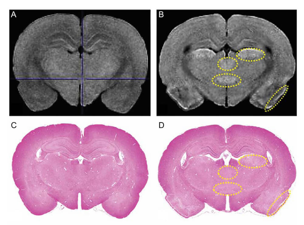
Preclinical noninvasive imaging can be an indispensable tool for studying animal models of disease. In vivo imaging to assess anatomical, functional, and molecular features requires verification by a comparison to the macroscopic and microscopic morphological features, since all noninvasive in vivo imaging methods have much lower resolution than standard histopathology. Comprehensive pathological evaluation of the animal model is underutilized; yet, many institutions have veterinary or human pathologists with necessary comparative pathology expertise. By performing a rigorous comparison to gross or histopathology for image interpretation, these trained individuals can assist scientists with the development of the animal model, experimental design, and evaluation of the in vivo imaging data. These imaging and pathology corroboration studies undoubtedly increase scientific rigor and reproducibility in descriptive and hypothesis-driven research. A review of case examples including ultrasound, nuclear, optical, and MRI is provided to illustrate how a wide range of imaging modalities data can be confirmed by gross or microscopic pathology. This image confirmation and authentication will improve characterization of the model and may contribute to decreasing costs and number of animals used and to more rapid translation from preclinical animal model to the clinic.
In Vivo Imaging With Confirmation by Histopathology for Increased Rigor and Reproducibility in Translational Research: A Review of Examples, Options, and Resources
View as PDF
Kathleen Gabrielson, Robert Maronpot, Sébastien Monette, Coraline Mlynarczyk, Yuval Ramot, Abraham Nyska, and Polina Sysa-Shah
View as PDF