Because the paired lobes (ventral, dorsal, lateral, and anterior) of the rat prostate have not been consistently sampled in many carcinogenicity and toxicity studies, comparison among different investigations has been compromised. The lack of specific site identification for prostatic lesions further lessens the value of incidences reported. We present here the lobe-specific incidences and degree of severity of background prostatic, seminal vesicular, and ampullary glandular lesions in 1768 control Fischer-344 rats from 35 recent National Toxicology Program 2-year carcinogenicity and toxicity studies conducted in 4 laboratories. The dorsal and lateral lobes were combined and considered the dorsolateral lobe where inflammation, epithelial degeneration, mucinous cysts, and edema were observed. Inflammation in the dorsolateral lobes was significantly associated with pituitary gland adenoma whose prolactin was suggested to play an important role in pathogenesis of prostatic inflammation. Epithelial degeneration, epithelial hyperplasia, inflammation, edema, and adenoma were conspicuous in the ventral lobes. Inflammation and edema occurred in the anterior lobes (coagulating glands). Inflammation, dilatation, epithelial hyperplasia, edema, and adenoma were observed in the seminal vesicles. Inflammation was also present in the ampullary glands. We suggest an optimal embedment and trimming method in rat prostate and seminal vesicle to ensure adequate, consistent sampling.
Keywords. Accessory male sex glands; background data; histopathology; rodents; pituitary gland adenoma; prolactin; spontaneous.
INTRODUCTION
Assessment of the potential toxicological and carcinogenic effects of a chemical compound in male accessory sex glands including prostate, seminal vesicle, and ampullary gland may be difficult if the incidence of spontaneous histopathological background lesions is unknown. Although the rat prostate has been used as an experimental model to analyze the function of androgenic hormones (26, 28), few background histopathological data exist for this gland and other male accessory sex glands in Fischer-344 (F-344) rats commonly used in 2-year carcinogenicity and toxicity studies (6, 11, 18,41,50).
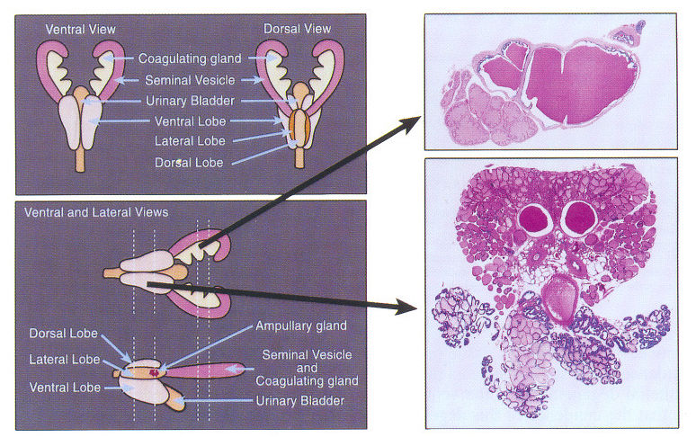
The rat prostate consists of 4 paired lobes including ventral, dorsal, lateral, and anterior (4, 23, 27, 28). The coagulating glands, which represent the anterior lobes, are easily distinguishable from the other prostatic lobes and have been reported separately from prostate in routine studies. Most biological studies of the rat prostate have been performed on the ventral lobe because gross visualization of the dorsal and lateral lobes is difficult (28). Although the ventral, dorsal, and lateral lobes differ morphologically and functionally (19, 20, 23, 28), few studies have identified these lobes separately (12, 13, 17, 18, 33-37, 50). Because the paired lobes of the rat prostate have not always been sampled consistently in past carcinogenicity and toxicity studies, comparison among different studies has been compromised. Omission of the identification of specific sites for prostatic lesions in controls lessens the value of reported background incidences. Histopathological lesions in the seminal vesicles and ampullary glands have seldom been reported (4,6-8, 18, 22, 30,46,50).
This retrospective histopathological examination was undertaken to document lobe-specific tissue accountability and incidences of background histopathological lesions in the prostate, seminal vesicles, and ampullary glands in control F-344 rats from 35 recent 2-year carcinogenicity and toxicity studies conducted in 4 laboratories for the National Toxicology Program (NTP). In addition, possible associations between various lesions were explored statistically; in particular, possible correlation between prostatic inflammation and the presence of pituitary gland adenomas was evaluated, because exposure to prolactin (PRL) has been implicated in the development of prostatitis (40, 42, 51, 52), and prolactinomas are common in F-344 rats.